Demo images from new MANUS scanner SW.
Recently released new software and firmware that open up exciting opportunities for using the latest techniques for even better ultrasound images, where you can see details that were not possible before.
Since much of the signal processing is done with powerful hardware/FPGA, we can create images where we combine the best of ultrasound techniques to achieve very high resolution images combined with high frame rate.
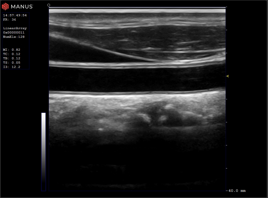
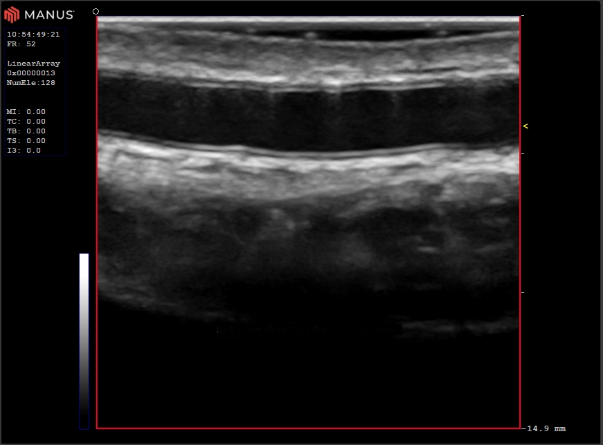
Carotid artery in longitudinal view.
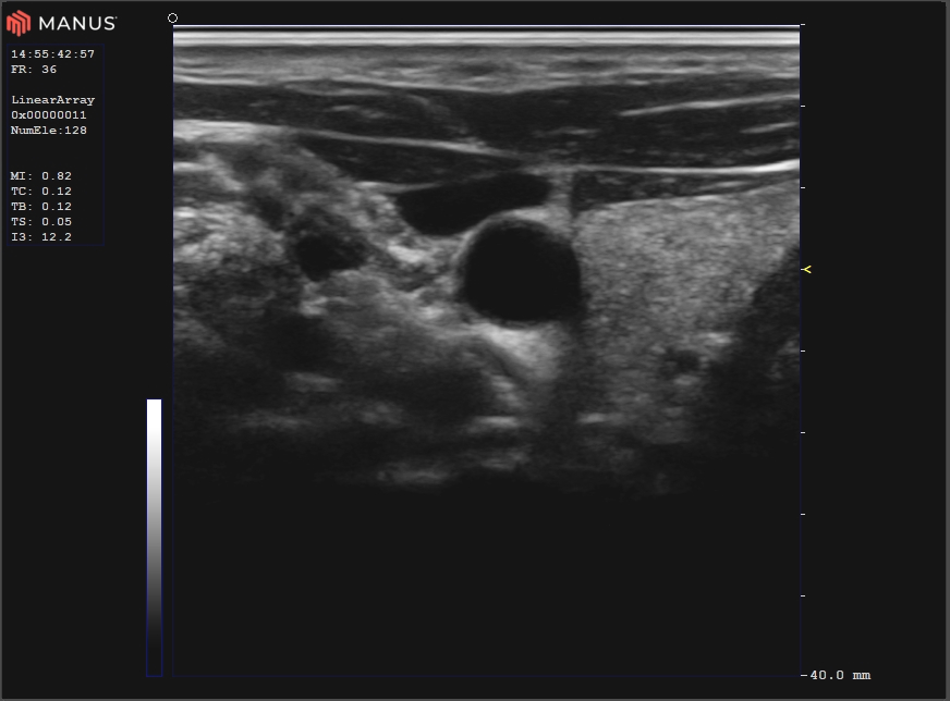
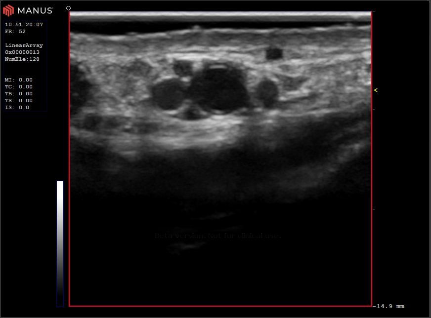
Carotid artery in sagital view with the thyriod gland.
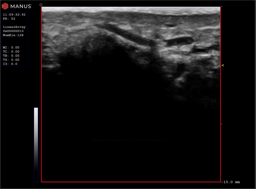
Artery in finger joint, Ø ~ 0.6 mm.
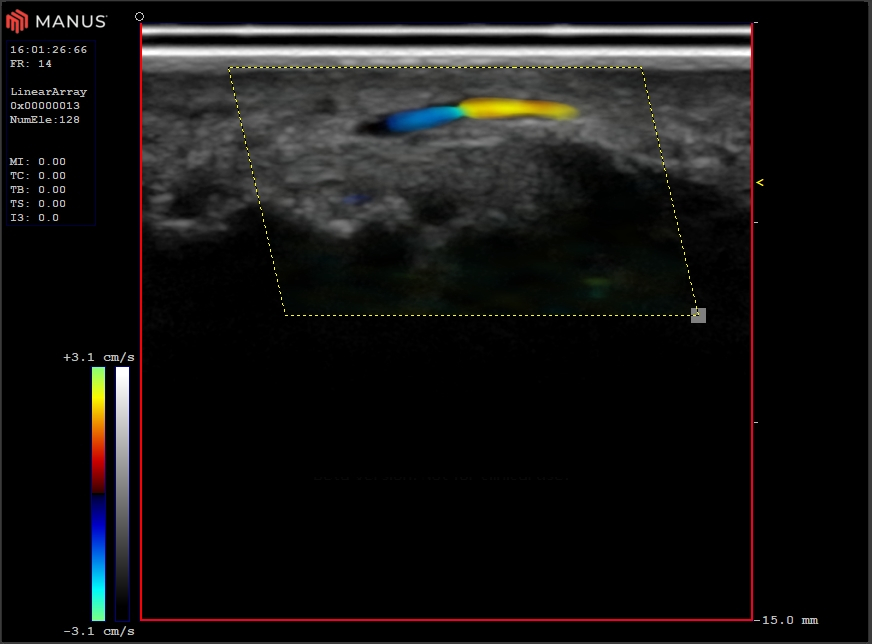
Finger tip vessel, Ø ~ 0.5 mm
Radial artery in longitudinal view.
Radial artery in sagital view.
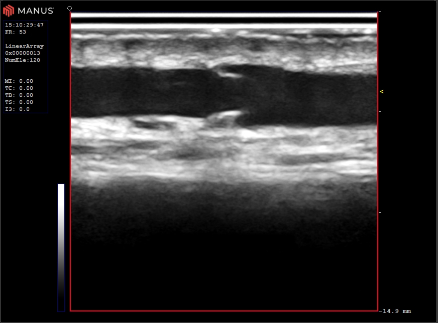
Vein valve.